-
 Trackback
-
 Orthopaedic appliance
-
 Chelonians
-
 Dermatome
-
 CryoSat
-
 Antitussive
-
 Mono-refringence
-
 Lander
-
 Ohm
Ohm
-
 A5
A5
-
 Kepler's laws
-
 Gravitation
-
 Limbic system
-
 Eczema
-
 Artificial CO2 sequestering
-
 Mercalli scale
-
 Olfactory centres
-
 Eutrophication
-
 Biosphere
-
 Bronchiole
-
 Typosquatting
-
 Marginal basin
-
 Triple therapy
-
 Abduction
-
 Supersymmetry
-
 Infusion
-
 Denial of service
-
 Gametogenesis
-
 Uracil
-
 Insulation
Radiography
Radiography is an imaging technique designed to visualise an organ or part of the body on a photosensitive film. It is performed by a hospital radiologist or in "primary care" and uses X rays. By extension, the term "radiograph" also describes the radiography film.
Radiography - the process
This investigation is used to visualise most organs. Bone, joint, or spinal radiography, pulmonary radiography, urinary or digestive radiography, PA (plain (unprepared) abdomen)… it has many areas of application.
The investigation procedure
In contrast to a CT scan, the radiograph involves using X rays. The beam is emitted from a fixed source (a tube) which does not rotate. The rays are absorbed by tissues to a greater or lesser extent depending on the density of the tissues and are then collected on a photosensitive film placed behind the patient. The X rays leave a greater or lesser opaque appearance on the film depending on the density of the tissues they pass through. In a specific investigation (for example the gastro-intestinal tract, or of a richly vascularised organ), and the doctor may initially decide to administer a contrast medium by injection. These can improve reading of the films.
Possible risks of radiography
A radiograph is not painful. Whilst it has no contraindications, some precautions must be taken, particularly in pregnant or breastfeeding women. You should discuss any concerns with your radiologist.
Source: Dictionnaire de l'imagerie médicale et des rayonnements, Académie nationale de Médecine, PUF
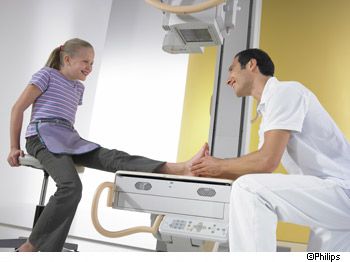
 Radiography involves the use of X rays. © Philips
Latest
Fill out my online form.


