-
 Typhoid fever
-
 CPT symmetry
-
 Fischer-Tropsch process
-
 DSL
DSL
-
 Peptide bond
-
 Complication
-
 Blende
-
 Ibuprofen
-
 Proof of security
-
 Submarine canyon
-
 Optoelectronics
-
 Eversion
-
 emf
emf
-
 Botulinum toxin
-
 Steppe
-
 Parabens
-
 Oxfam
Oxfam
-
 Henry
Henry
-
 Top plate
-
 Raspberry
-
 Linoleic acid
-
 Sun
Sun
-
 Ampholyte
-
 Dwarf galaxy
-
 Measles
-
 Extrasolar
-
 Playlist
-
 Unsanitary water
-
 Eclogite
-
 Silicate
Light microscope
The light microscope visualises objects or details which are invisible to the eye, which does not have sufficient resolution to see them.
Technique
The light microscope uses light. It has two lenses:
- a magnifying lens, to magnify the object to be looked at (several magnifications are available);
- and the ocular lens so that the light rays arrive in parallel at theeye allowing the eye to rest.
Additional instruments are used to adjust the amount of light (diaphragm) or focus (knobs connected to a focusing system) in order to increase the detail of the sample on the sample-holding slide.
The resolution of light microscopes is up to 0.2 micrometres and is limited by diffraction of light. There are methods available that come close to this limit by using an immersion lens (in oil) or by reducing the light wavelength (although this is restricted to the visible spectrum).
Use of the light microscope
The light microscope can visualise the individual cells of living objects (bacteria, yeasts, unicellular organisms) or fixed objects (tissue sections). It is also used to study the physics of materials and in geology.
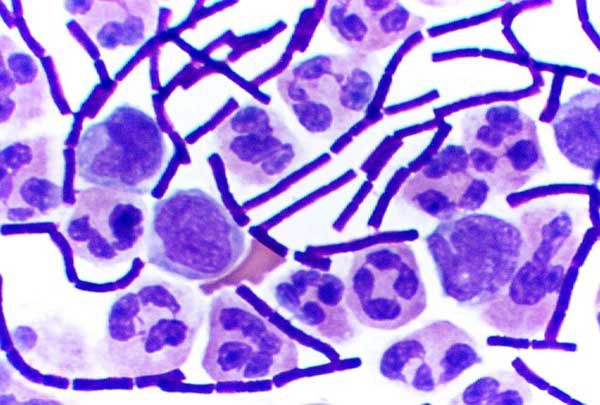
The objects become very clear under the illumination although the tissues often need to be stained in order to examine them. As not all tissues take up the stain in the same way these staining techniques can also be used to distinguish specific objects or to differentiate between two similar organisms (Gram staining for bacteria).
Many variants of light microscopy are now used (phase contrast, dark background, polarised light, fluorescence, confocal etc.).
 The Gram stain can distinguish between the two main types of bacteria (Gram positive and Gram negative). The rods are Bacillus anthracisbacteria. The other cells belong to the immune system. © DR
Latest
Fill out my online form.


