-
 Diorite
-
 Postulate
-
 NEAR-Shoemaker
-
 Single-stranded
-
 Feral
Feral
-
 Immunoglobulin
-
 Green flash
-
 ISDB-T
-
 Threshold dose
-
 Gustatory
-
 General relativity
-
 Bartholin glands
-
 Thyroid
-
 Predisposition
-
 Electrical resistance
-
 Phonolite
-
 Naevus
-
 Heavy mud
-
 Feather
-
 Breast-shield
-
 Wild cherry
-
 Pearlite
-
 London plane tree
-
 Epiphysis
-
 Truncation
-
 Villi
Villi
-
 Electrocution
-
 Maser
Maser
-
 Osteoarthritis
-
 Incandescence
Endoscopy
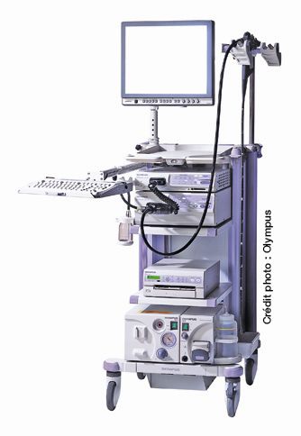
An endoscopy is an investigation that visualises the inside of organs, a natural channel or a cavity. The investigation is performed with a rigid or more commonly a flexible tube, fitted with optical fibres and a light source. It is linked to a video camera and displays the images obtained on a screen, as it passes through the body. The investigation is either performed in hospital or in specialist consulting rooms.
Endoscopy practice
Endoscopy is either used to make a diagnosis or to treat a disease. The scope of endoscopy has increased due to advances in the materials used. The endoscope is inserted through a natural track: the mouth when the stomach or bronchi are to be examined, the nostrils for nasal fossae, vocal chords and sinuses, and the anus to examine the colon. Small incisions may be necessary - for example in the abdomen - for other investigations. There are several types of endoscopy:
- arthroscopy,
- colonoscopy,
- rectoscopy,
- hysteroscopy, and
- pleuroscopy
A sample (a biopsy) can be taken by using guided forceps fixed to the end of an endoscope. An endoscope equipped with ultrasound probes can also be used to measure blood flow or to produce an image of certain lesions.
The procedure for endoscopy
Depending on the organ or the cavity being investigated, local or general anaesthesia may be offered. In these cases, a pre-operative visit with an anaesthetist is obligatory. Patients may be hospitalised for some investigations, for several days after the investigation so that they can be observed.
Possible risks of endoscopy
Each type of endoscopy has its own risks. However; the risks are generally mostly those resulting from infection caused by unhygienic probe or instrument cleaning processes. The possible risks of any anaesthetic must also be considered.
Source: Merck Manual (fourth edition), Gustave Roussy Institute website, accessed 7 January 2011
 Endoscopy: a close-up on life of organs © Olympus
Latest
Fill out my online form.


