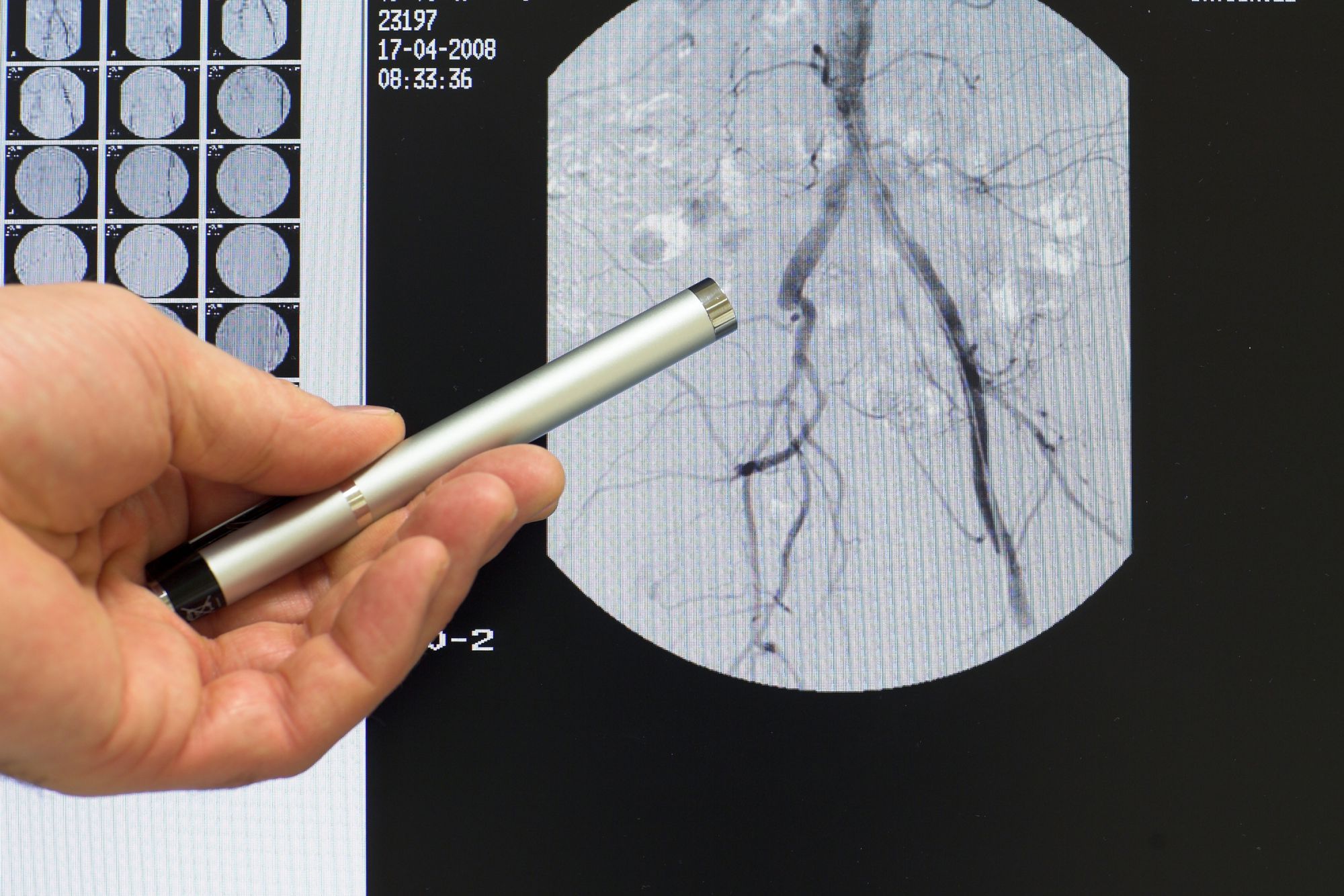
Angiography
Angiography is used to examine the inside of blood vessels. Specialists refer toarteriography when the examination specifically targets an artery. In contrast, they refer tophlebography (or venography) for a vein. Angiography is always performed by a radiologist.
Angiography - the process
This type of examination is normally indicated when a blood vessel disease or an arterial or venous malformation, is suspected. It can also reveal narrowing or obstruction of an artery or vein.
The examination process
Angiography is performed under local or general anaesthesia depending on the case. Arteriography begins by puncturing an artery. This is usually the carotid (in the neck), the humeral artery (in the elbow) or the femoral artery (in the groin) depending on the area which the investigator is looking at in the brain, heart, eye or lung, etc. The doctor introduces a catheter that is a flexible tube that he/she advances while monitoring its progress on a control screen. The instrument is used to inject an iodinated contrast medium that colours the vessels. Once the examination has been completed and the radiology films have been taken, the catheter is removed and the puncture site is compressed for a few minutes.
Possible risks of angiography
Apart from local reaction at the puncture site (such as haematoma formation) the main risk is from an intolerance reaction to the iodinated compound. Consequently, allergic patients need to tell their doctor about any past history. The risk of thrombo-embolism from advancing the catheter is very rare and believed to be less than 1%.
Source: Société française de radiologie
 Angiography is performed by a radiologist. © Bergringfoto/Fotolia
Latest
Fill out my online form.


